Upper Thigh Muscles Ct Anatomy / Figure 6 from Normal MR imaging anatomy of the thigh and ... : It crosses the back of the shoulder and attaches to the upper humeral shaft, below the head.
Upper Thigh Muscles Ct Anatomy / Figure 6 from Normal MR imaging anatomy of the thigh and ... : It crosses the back of the shoulder and attaches to the upper humeral shaft, below the head.. Muscles of the posterior cervical and upper thoracic spine 1. One of those muscles, the psoas major, is also important for posture Superior ramus of the pubis insertion: It has a dual innervation, and thus can be considered a transitional. Thigh muscle strains are common for people of all ages.
To better understand how to best target the arm musculature, let's first delve into basic anatomy. It arises by tendinous fibers from the anterior superior iliac spine and the upper half of the notch below it. Muscles are named according to their shape, location, or a combination. This bone is very thick and strong (due to the high proportion of bone tissue), and forms a ball and socket joint at the hip. The chewing muscles enable you to chew your food by moving the upper and lower teeth against one another.
Muscles of the Leg (Human) from anatomycorner.com In human anatomy, the thigh is the area between the hip (pelvis) and the knee. In the upper back region, the trapezius, rhomboid major, and levator scapulae muscles anchor the scapula and clavicle to the spines of several vertebrae and in addition to moving the arm and pectoral girdle, muscles of the chest and upper back work together as a group to support the vital process of. Thigh muscle strains are common for people of all ages. It arises by tendinous fibers from the anterior superior iliac spine and the upper half of the notch below it. Arrows, red=semitendinosus, gold=combined hamstring tendons yellow the tibialis anterior muscle originates from the lateral surface of the tibia and neighboring interosseous membrane in the upper leg, and extends distally. 2, vastus medialis & intermedius muscles. Anterior muscles extend your legs and flex your thighs. The pectineus muscle is a flat muscle that forms the base of the femoral triangle.
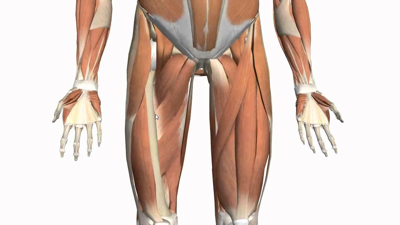
The muscles of the thigh are arranged into three compartments.
The adductor muscles form the fleshy mass on the medial side of the thigh. It has a dual innervation, and thus can be considered a transitional. In clinical anatomy the thigh muscles are divided into three groups: Muscles are named according to their shape, location, or a combination. Lesser trochanter to linea aspera nerve supply:( double nerve. You've got an anterior compartment, medial, and posterior compartment and these are separated by the intermuscular this is this group of muscles here anteriorly in the thigh, obviously and these muscles are supplied by the femoral nerve. As the cursor is moved over a particular compartment of the lower. Learn faster with these free muscle labeling diagrams. Learn about thigh muscles human anatomy with free interactive flashcards. This is my video about the muscles of the back. Pain in the upper thighlearn about different causes of upper thigh pain, from injuries to nerve problems. Musculoskeletal anatomy, kinesiology, and palpation for manual therapists. Anatomical structures of the lower limb (hip, thigh, knee, leg, ankle and foot) and specific regions (compartment of the lower limb) are visible on cross section of the leg :
2, vastus medialis & intermedius muscles. Pain in the upper thigh can be difficult to diagnose because this area of the body contains many muscles, tendons, and ligaments. This injury frequently occurs near the point where the. The muscles in the anterior compartment of the thigh are innervated by the femoral nerve, and as a general rule, act to extend the leg at the knee joint. The sparthos thigh compression sleeve provides compression as well as support for thigh muscles.
Muscles of the Thigh and Gluteal Region - Part 2 - Anatomy ... from i.ytimg.com The adductor muscles form the fleshy mass on the medial side of the thigh. We look at the associated symptoms and treatment options. The sparthos thigh compression sleeve provides compression as well as support for thigh muscles. In clinical anatomy the thigh muscles are divided into three groups: It has a dual innervation, and thus can be considered a transitional. It is part of the lower limb. Anatomy of the human body. One of those muscles, the psoas major, is also important for posture
Anterior muscles extend your legs and flex your thighs.
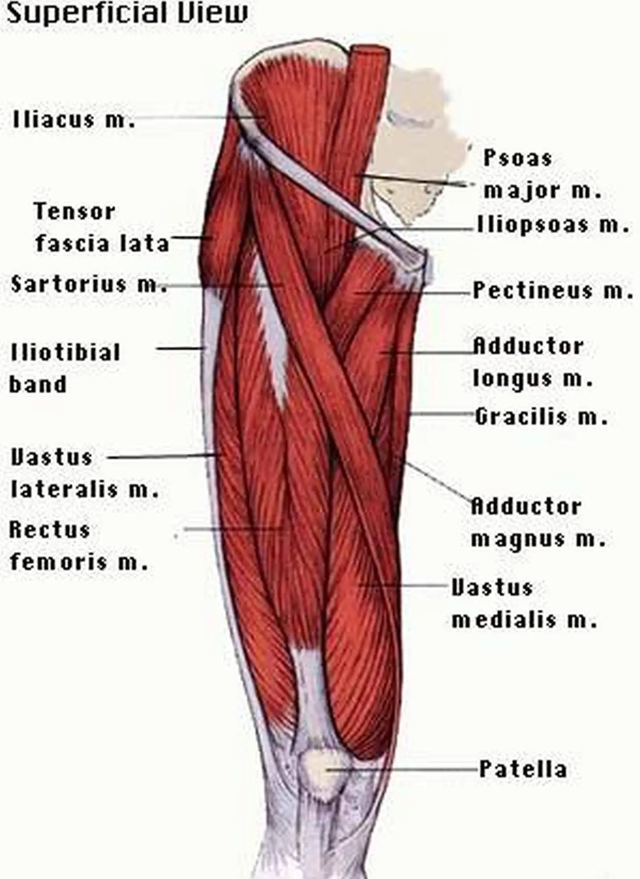
Muscle anatomy of upper thigh, human muscles, muscle anatomy of upper thigh. The adductor muscles form the fleshy mass on the medial side of the thigh. Create flashcards for free and quiz if you like muscles of upper limb, you might love these ideas. The thigh has three sets of strong muscles: Reviewed by mary rodts, dnp. To better understand how to best target the arm musculature, let's first delve into basic anatomy. It crosses the back of the shoulder and attaches to the upper humeral shaft, below the head. Case contributed by dr roberto schubert. Simple and easy notes for quick revision. In clinical anatomy the thigh muscles are divided into three groups: The iliopsoas is made up of two muscles that flex the thigh. Want to learn more about it? The thigh is the area between the hip and the knee joint.
Muscular compartment, bones (tibia, fibula) and muscles. Lesser trochanter to linea aspera nerve supply:( double nerve. It has a dual innervation, and thus can be considered a transitional. Want to learn more about it? Pain in the upper thighlearn about different causes of upper thigh pain, from injuries to nerve problems.
Pictures Of Anterior Thigh Muscles from healthiack.com Learn about thigh muscles human anatomy with free interactive flashcards. Thigh muscle strains can occur when playing sports or participating in a daily activity. This webpage presents the anatomical structures found on thigh mri. The muscles that move the forearm are located along the humerus, which include the triceps brachii, biceps brachii, brachialis, and brachioradialis. When a muscle is stretched beyond its limit, a tear can occur that can range from mild to serious. 2, vastus medialis & intermedius muscles. Muscles of the posterior cervical and upper thoracic spine 1. In the upper back region, the trapezius, rhomboid major, and levator scapulae muscles anchor the scapula and clavicle to the spines of several vertebrae and in addition to moving the arm and pectoral girdle, muscles of the chest and upper back work together as a group to support the vital process of.
Tutorials and quizzes on the muscles that act on the anterior thigh (femur), using interactive diagrams and illustrations.
One of those muscles, the psoas major, is also important for posture The sparthos thigh compression sleeve provides compression as well as support for thigh muscles. Muscles that move the shoulder and arm include the trapezius and serratus anterior. 2, vastus medialis & intermedius muscles. Thigh muscle strains are common for people of all ages. This injury frequently occurs near the point where the. It has a dual innervation, and thus can be considered a transitional. Muscles are named according to their shape, location, or a combination. In human anatomy, the thigh is the area between the hip (pelvis) and the knee. Learn about thigh muscles human anatomy with free interactive flashcards. The hamstring muscles in the back of the thigh, the quadriceps muscles in the front, and the muscle strains usually happen when a muscle is stretched beyond its limit, tearing the muscle fibers. The posterior compartment of the thigh contains the knee flexors and hip extensors.it has the following muscles, nerves and vessels: Musculoskeletal anatomy, kinesiology, and palpation for manual therapists.
The hamstring muscles in the back of the thigh, the quadriceps muscles in the front, and the muscle strains usually happen when a muscle is stretched beyond its limit, tearing the muscle fibers upper thigh anatomy. Muscles of the posterior cervical and upper thoracic spine 1.